
Hyperkalemia ECG Case: A Slow and Fatal Presentation with Bradycardia and Sine Waves
Case Presentation (H2) A 69-year-old male patient presented to the Emergency Department with: Bradycardia
A Slow & Fatal ECG Pattern: Severe Hyperkalemia
Case Presentation
A 69-year-old male patient presented to the Emergency Department with:
- Bradycardia
- Hypotension
- General appearance of being acutely unwell
No prior medical history was available at presentation.
The ECG obtained revealed a critical pattern.
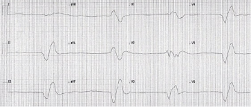
What Is Your Diagnosis and How Would You Manage This?
Let’s analyze the ECG step-by-step.
Why This Case Is Important
This case is valuable for several reasons:
- It represents a common life-threatening emergency
- It is potentially reversible with proper intervention
- It gives us an opportunity to learn both diagnosis and management
ECG Features Observed
- Bradycardia
- Very broad QRS complexes
- Irregular rhythm
- No visible P waves
- Hyperacute (tall) T waves
- Sine wave morphology
Diagnosis: Severe Hyperkalemia
This was a case of Hyperkalemia, confirmed with a potassium level of 10.5 mmol/L.
ECG Manifestations of Hyperkalemia
Although hyperkalemia can present with a wide range of ECG abnormalities, typical features include:
- Flattened or lost P waves
- Prolonged PR interval
- Widened QRS complex
- Patterns that mimic ventricular tachycardia (VT)
- ST segment elevation (especially in V1–V2)
- Hyperacute T waves
- Arrhythmias (e.g., AV blocks, bradycardia)
- Pseudo-myocardial infarction patterns (e.g., new LBBB + ST elevation)
- Sine waves
Important: Misdiagnosing hyperkalemia as VT and treating with antiarrhythmics can be fatal.
Additional ECG Examples
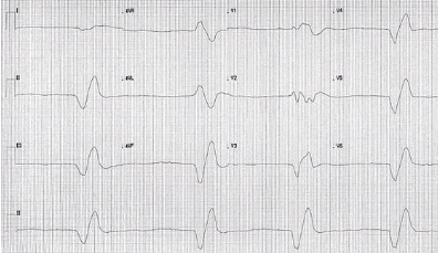
Sine wave appearance with K+ of 8.2 mmol/L
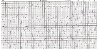
Broad complex tachycardia mimicking VT at K+ 7.8 mmol/L
Reversible cases after correct treatment
Treatment of Severe Hyperkalemia
1. IV Calcium – First Line Treatment
Calcium Chloride 10% – 10 mL IV
- Pros: 3x stronger than calcium gluconate, effective immediately
- Cons: Irritant to veins; requires central line or large peripheral vein
Calcium Gluconate 10% – 30 mL IV
- Pros: Safer via peripheral access
- Cons: Weaker; slower effect; requires hepatic metabolism
Repeat after 5 minutes if needed.
2. Beta-2 Agonist Nebulizer
- Salbutamol 10–20 mg nebulized
3. Insulin + Glucose
- 25 g Glucose + 10 units IV soluble insulin
- Give over 15 minutes
4. Sodium Bicarbonate (NaHCO₃)
- Reserved for peri-arrest situations with metabolic acidosis or renal failure
- Not routinely used unless indicated (per UK Resuscitation Council)
Management Algorithm
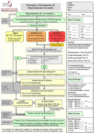
Refer to this Resuscitation Council UK algorithm for a practical summary.
Author note: There’s a newer version, but this one remains more intuitive.
Outcome
Despite full administration of the hyperkalemia protocol, the patient unfortunately went into cardiac arrest.
Return of Spontaneous Circulation (ROSC) could not be achieved.
Further Reading
Key Takeaways
- Hyperkalemia is a deadly yet reversible condition
- Sine wave ECG + broad QRS + tall T waves = treat immediately
- Always treat the electrolyte, not the rhythm
- Misinterpreting hyperkalemia as VT can be fatal
FAQ – A Slow & Fatal ECG Pattern
Q1: What are the ECG signs of severe hyperkalemia?
Key findings include bradycardia, absent P waves, broad QRS complexes, tall T waves, sine wave morphology, and arrhythmias.
Q2: What is the first-line treatment for hyperkalemia with ECG changes?
Intravenous calcium (10 mL CaCl₂ or 30 mL Ca gluconate) is given immediately to stabilize cardiac membranes.
Q3: Can hyperkalemia mimic ventricular tachycardia?
Yes. Broad complex rhythms without P waves can mimic VT. Treating as VT instead of correcting potassium can be fatal.
Ready To Take Your Clinical Learning Further?
Explore structured ECG and emergency medicine courses built around practical clinical knowledge, real cases, and confident decision-making.