
Fast & Furious ECG Series – Part 1: Atrial Flutter vs SVT
Case Presentation (H2) A 53-year-old male presented to the Emergency Department with palpitations. His ECG showed a heart rate of 132 bpm.

Fast & Furious (Part 1): Narrow Complex Regular Tachycardia
Case Presentation
A 95-year-old woman presented to the Emergency Department with complaints of palpitations.
Her heart rate was 144 bpm.
An ECG was obtained, revealing a narrow-complex regular tachycardia.
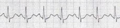
What Is Your Diagnosis?
And why?
Narrow Complex Regular Tachycardia: Differential Diagnosis
This ECG represents a classic theme seen in many emergency cases: narrow-complex regular tachycardia.
The most common differentials include:
-
Sinus Tachycardia
- Clear, single P wave before every QRS complex
-
Supraventricular Tachycardia (SVT)
- Typically no clear P waves visible
-
Atrial Flutter with Fixed Block
- Multiple P waves (flutter waves) before each QRS complex
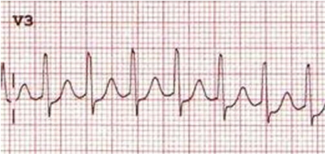
Visual ECG examples can help distinguish these — but in some real-world cases like this one, they aren’t always reliable.
Why This Case Is Tricky
Initially, there appeared to be only one visible wave between each QRS complex.
That could suggest:
- P waves → Possibly Sinus Tachycardia with 1st degree AV block
- T waves → Possibly SVT
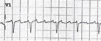
So which is it? Diagnosis is critical — the treatment paths are entirely different.
ECG Analysis
Clue #1: Heart Rate
- 144 bpm is relatively slow for SVT
- But it's just right for Atrial Flutter (classically around 150 bpm)
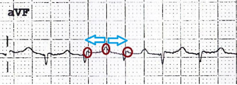
Clue #2: The “Bix Rule”
When you see a “P wave” exactly midway between two QRS complexes, assume there are two more flutter waves — one buried before and one after the QRS.
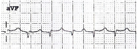
In this case, the “single wave” seen between QRS complexes was actually part of a flutter wave series.
Final Diagnosis: Atrial Flutter
- Atrial flutter with a fixed 2:1 block was the working diagnosis.
- The patient was allergic to beta-blockers, so she was treated with Verapamil (CCB).
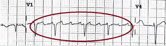
- Post-treatment ECG revealed classic flutter waves in V1 with variable block.
Learning Points
- Always consider atrial flutter when the heart rate is close to 150 bpm
-
Our brains are often trained to ignore inverted P waves, misreading them as T waves
- Try flipping the ECG upside down when in doubt
-
Use the Bix Rule
- If a “P wave” is seen midway between QRS complexes, suspect hidden flutter waves
- The differential for narrow complex regular tachycardia = Sinus tachycardia, SVT, or Atrial Flutter with fixed block
Further Learning
Watch the full case discussion on YouTube
Key Takeaways
- Not all regular tachycardias are SVT — look deeper for flutter
- Heart rate near 150 is a huge clue for atrial flutter
- Inverted P waves ≠ T waves — flip the ECG when in doubt
- Clinical decisions depend on accurate rhythm recognition
FAQ – Fast & Furious (Part 1)
Q1: What are the most common causes of narrow-complex regular tachycardia?
The key differentials include sinus tachycardia, supraventricular tachycardia (SVT), and atrial flutter with fixed block.
Q2: How can I identify atrial flutter on ECG if flutter waves are not obvious?
Use the Bix Rule — if a P-like wave is exactly between QRS complexes, suspect hidden flutter waves before and after.
Q3: Why is heart rate important in differentiating SVT from atrial flutter?
A heart rate close to 150 bpm is highly suggestive of atrial flutter with 2:1 block, especially if the rhythm is regular.


Ready To Take Your Clinical Learning Further?
Explore structured ECG and emergency medicine courses built around practical clinical knowledge, real cases, and confident decision-making.