
A Cold Case: ECG Clues Unveil Hidden Hypothermia
Case Presentation (H2) A 46-year-old male patient with a high BMI and a history of paraplegia secondary to an old spinal cord injury was discovered on the floor by his carer in the early morning. It’s unclear how long he remained there, but it was likely overnight. Upon assessment, the patient appea

Case Presentation

A 46-year-old male patient with a high BMI and a history of paraplegia secondary to an old spinal cord injury was discovered on the floor by his carer in the early morning.
It's unclear how long he remained there, but it was likely overnight.
Upon assessment, the patient appeared confused, though hemodynamically stable.
Blood work revealed Acute Kidney Injury (AKI), likely due to rhabdomyolysis from prolonged immobility.
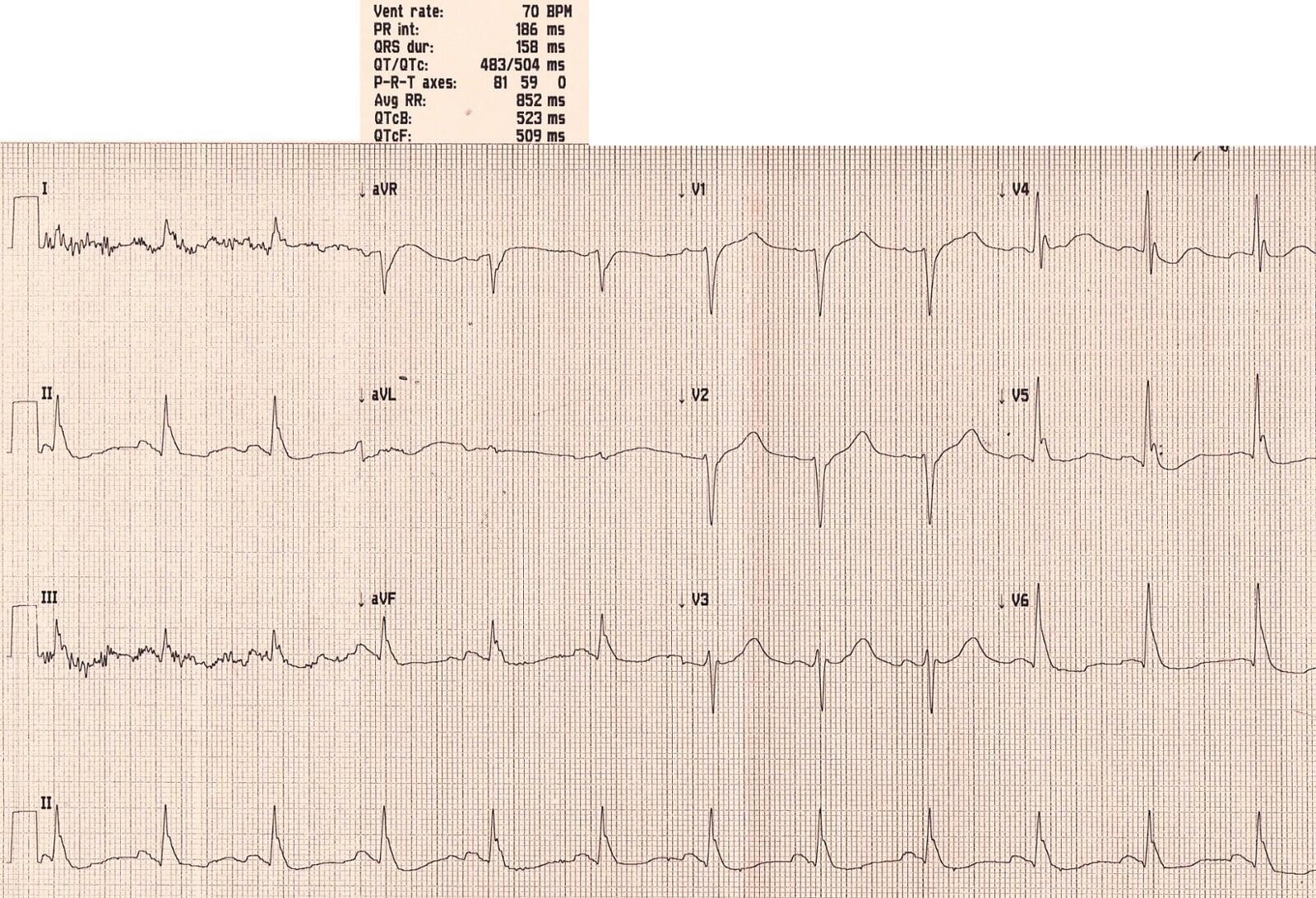
Initial ECG Findings
Can you guess what caused the ECG abnormalities?
1. Prolonged QTc
QTc = 523 ms.
This is a critical red flag. A prolonged QTc can have several causes:
Causes of QTc Prolongation
Congenital:
- Romano-Ward syndrome
Metabolic (All the Hypo's):
- Hypokalemia (K)
- Hypocalcemia (Ca)
- Hypomagnesemia (Mg)
- Hypothermia
- Hypothyroidism
Medications:
- Tricyclic antidepressants (TCA)
- Amiodarone
- Antipsychotics
- Antidepressants
- Antihistamines
- Ondansetron
- Macrolides (e.g., Clarithromycin)
- Methadone
Other causes:
- Increased Intracranial Pressure (ICP)
- Acute Coronary Syndrome (ACS)
A Common ED Trap
Imagine a young patient with mental health conditions presenting to the ED with a chest infection and nausea.
They receive Amoxicillin + Clarithromycin and Ondansetron — a recipe for disaster if they're already on antidepressants or antipsychotics.
Without an ECG, a dangerously prolonged QTc could go unnoticed, putting the patient at serious risk for ventricular tachycardia (VT).
Osborn Waves
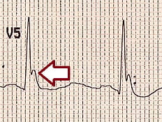
A distinctive wave at the end of the ECG complex stood out:
- Positive in positive complexes
- Negative in negative complexes
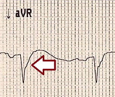
Known as Osborn Waves, these occur as deflections at the J-point, most visible in leads V2–V6, and are classically linked to:
- Hypothermia
- Hypercalcemia
- Raised ICP
Shivering Artifacts
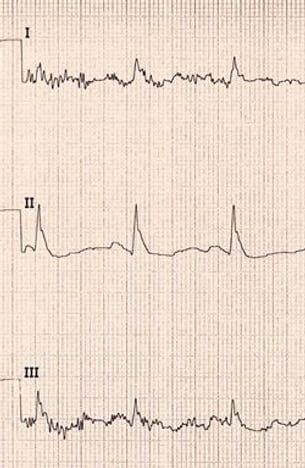
Movement artifacts were noted in leads I and III, consistent with shivering.
Diagnosis: Hypothermia
The diagnosis was based on the combination of:
- QTc prolongation
- Osborn waves
- Shivering artifacts
The likely diagnosis: Hypothermia.
This was confirmed by a core body temperature of 29°C, following prolonged exposure due to being immobilized overnight.
ECG should not replace core temperature measurement, but can provide valuable diagnostic clues in emergency settings.
ECG Changes in Hypothermia
- Prolonged PR, QRS & QT intervals
- Bradyarrhythmias (e.g., sinus bradycardia, slow AF)
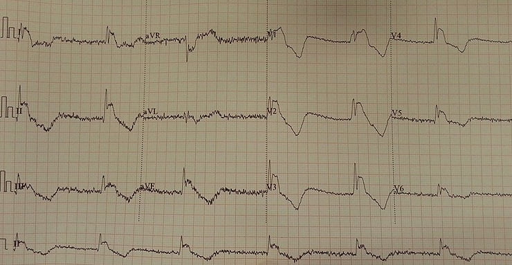
- Osborn (J) waves — may mimic STEMI
- Shivering artifacts
- Ventricular ectopics
Additional ECG Examples
- A hypothermic patient mimicking STEMI
- Temperature at 28°C with all hallmark findings
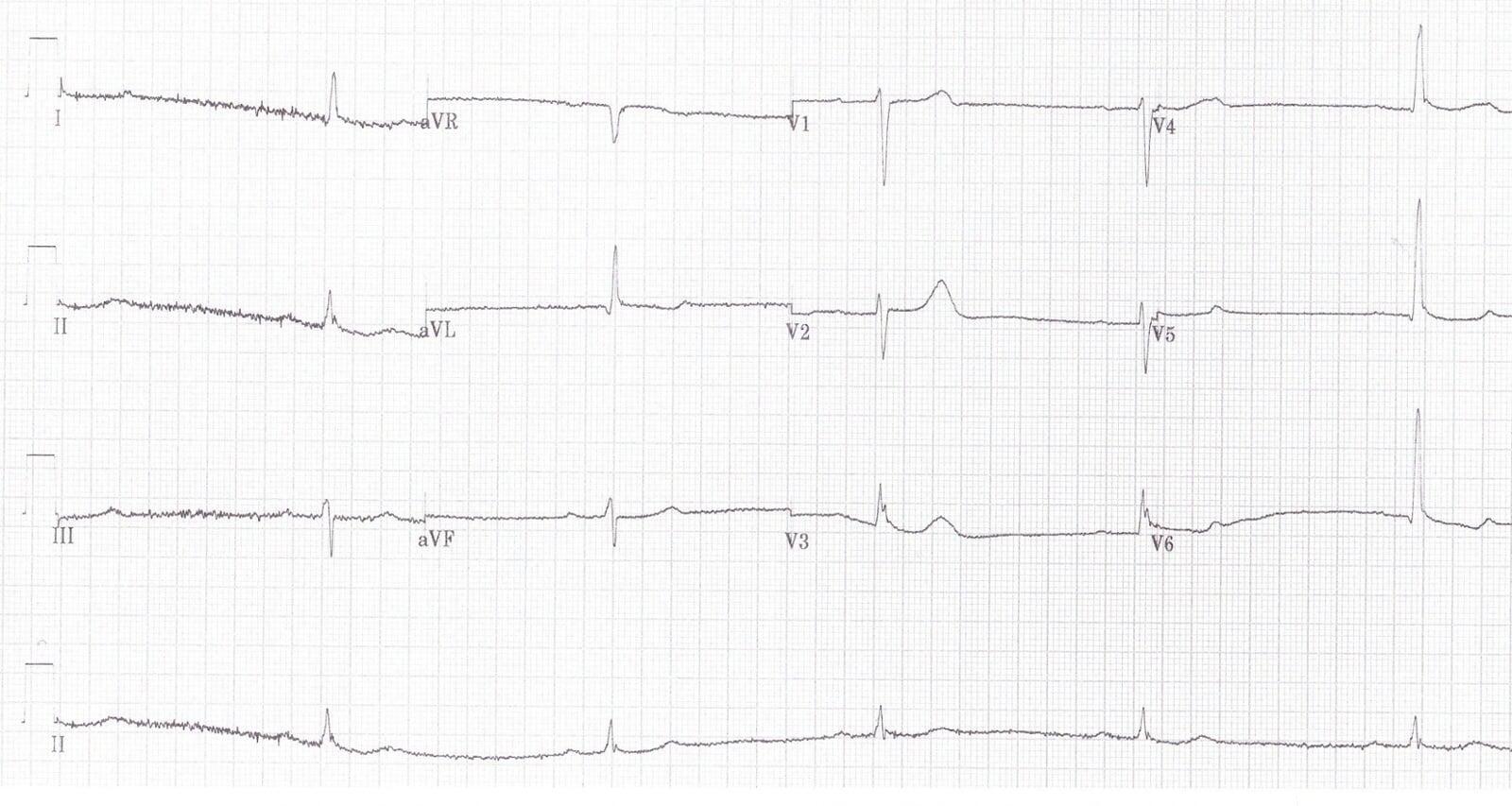
Case courtesy: Tom Cromwell, UK.
Further Reading
- Hypothermia ECG Library – Life in the Fast Lane (LITFL)
Key Takeaways
- Always consider hypothermia in confused patients with prolonged QTc and Osborn waves.
- Be vigilant about QTc-prolonging drug combinations in emergency care.
- ECG remains a critical, rapid, and informative tool for diagnosing underlying systemic conditions.
Frequently Asked Questions
What are the ECG signs of hypothermia?
ECG signs of hypothermia include prolonged PR, QRS, and QT intervals, Osborn waves

Ready To Take Your Clinical Learning Further?
Explore structured ECG and emergency medicine courses built around practical clinical knowledge, real cases, and confident decision-making.