
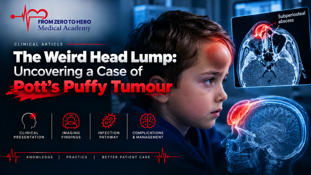
The Weird Head Lump: Uncovering a Case of Pott’s Puffy Tumour
Case Presentation (H2) A 69-year-old male presented to the Emergency Department feeling unwell, confused, and complaining of a forehead swelling that developed over one week, worsening significantly in the last 24 hours. Past Medical History:
Case Presentation
A 69-year-old male presented to the Emergency Department feeling unwell, confused, and complaining of a forehead swelling that developed over one week, worsening significantly in the last 24 hours.
Past Medical History
- Recurrent sinusitis.
Observations in ED
- Pulse: 125 bpm
- Blood Pressure: 95/55 mmHg
- Respiratory Rate: 28 bpm
- Oxygen Saturation: 94% on room air
- GCS: 13/15
A photo of the forehead lump was obtained with the patient’s permission.

What Is the Most Likely Diagnosis?
Given the clinical presentation, location, and shape of the swelling, the most likely diagnosis is: Pott’s Puffy Tumour
Understanding Pott’s Puffy Tumour
What is Pott’s Puffy Tumour?
- A rare, serious complication of frontal sinusitis or facial trauma.
- Represents osteomyelitis of the frontal bone with an associated subperiosteal abscess.
- First described by Sir Percival Pott in 1768.
Clinical Presentation
Typical symptoms include:
- Headache
- Periorbital swelling
- Fever
- Purulent rhinorrhoea
- Vomiting
Neurological signs such as meningitis or encephalitis can occur if the infection spreads intracranially.
Complications
Pott’s Puffy Tumour may lead to:
- Meningitis
- Epidural abscess
- Subdural abscess
- Venous sinus thrombosis
- Orbital cellulitis or intra-orbital abscess (if infection spreads downward)
Investigations
- CT scan or MRI of the head is critical for diagnosis.
- Misdiagnosing it as a simple infected cyst can be fatal.
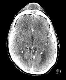
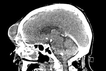
The CT scan of this patient, obtained with permission, confirmed the diagnosis.
Treatment
- Surgical drainage of the abscess is the standard of care.
- Broad-spectrum intravenous antibiotics covering gram-positive organisms and anaerobes are recommended for 4–6 weeks.
Further Learning
Watch the detailed case discussion on YouTube
Key Takeaways
- Pott’s Puffy Tumour is a rare but life-threatening complication of sinusitis.
- Early imaging is crucial to differentiate it from benign conditions like skin cysts.
- Prompt surgical and medical treatment are necessary to prevent serious complications.
Stay tuned for more fascinating radiology cases!
FAQ
Q1: What is Pott’s Puffy Tumour?
It is a frontal bone osteomyelitis with a subperiosteal abscess, often following frontal sinusitis or trauma.
Q2: What are the complications of Pott’s Puffy Tumour?
Complications include meningitis, epidural abscess, subdural abscess, venous sinus thrombosis, and orbital infections.
Q3: How is Pott’s Puffy Tumour diagnosed and managed?
Diagnosis is through CT or MRI imaging, and management involves surgical drainage plus 4–6 weeks of broad-spectrum antibiotic therapy.



Ready To Take Your Clinical Learning Further?
Explore structured ECG and emergency medicine courses built around practical clinical knowledge, real cases, and confident decision-making.