
My Chest Hurts – But It’s Not Pericarditis: ECG Clues to Pneumothorax
Case Presentation: A 21-year-old male presented to the Emergency Department with vague right-sided chest pain for several hours. He was normally healthy
Case Presentation
A 21-year-old male presented to the Emergency Department with vague right-sided chest pain for several hours.
- He was normally healthy.
- Vitals: Mild tachycardia, otherwise normal.
- No other clinical information was initially available.
An ECG was performed.
What’s the differential diagnosis?
How do you approach this when information is limited?
Let’s break it down the same way it unfolded in real time.
Initial Clinical Impression
This case reflects what ED doctors often experience:
A young patient + non-specific chest pain + ST elevation on ECG + limited info.
A nurse hands you the ECG before you've seen the patient — sound familiar?
Step 1: Build the Differential
Possible diagnoses:
- STEMI
- Pericarditis
- Benign Early Repolarization (BER)
- Pneumonia
- Pulmonary Embolism (PE)
- Aortic Dissection
- Pneumothorax
Step 2: Rule Out What You Can
Ruled out quickly:
- Pneumonia – Doesn’t fit clinically or on ECG.
- PE – Ruled out via Wells' + PERC scores.
- BER – Ruled out via old ECG showing no ST elevation.
- STEMI – Unlikely, but not impossible in young patients.
- Aortic dissection – Planned workup with chest X-ray, echo, and D-dimer.
Now we’re left with:
- Pericarditis
- Pneumothorax
ECG Interpretation
The ECG suggested acute pericarditis, with:
- Diffuse saddle-shaped ST elevation
- Spodick's sign
- PR elevation in aVR
- PR depression in other leads
Diagnosis at this point: Acute Pericarditis.
But then came the surprise.
Chest X-ray Changed Everything
CXR revealed a right-sided pneumothorax.
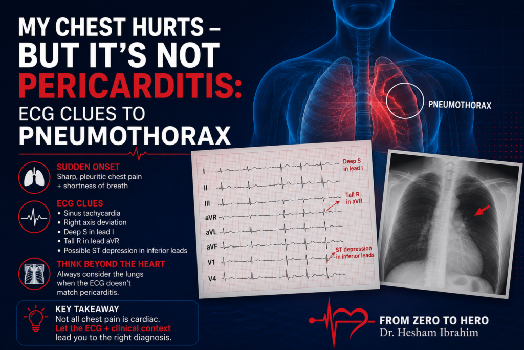
ECG Changes in Pneumothorax
Although underreported in literature, here are the summarized ECG findings:
Left-sided Pneumothorax
- Right axis deviation
- Loss of R wave progression (V1–V6)
- Low voltage
- T wave inversion (V1–V6)
- PR + ST segment changes
Right-sided Pneumothorax
- Loss of R wave progression (V1–V6)
- Q waves in inferior leads
- ST elevation (but no ST depression reported)
Mechanisms Behind ECG Changes
- Cardiac displacement or rotation
- Right ventricular strain/dilation
- Air insulation between heart and electrodes (low voltage)
Final Diagnosis: Right-Sided Pneumothorax
Despite an ECG mimicking pericarditis, the actual diagnosis was right-sided pneumothorax.
This case is a reminder to always consider pneumothorax in patients with unexplained chest pain and ST elevation.
Key Learning Points
- ECG changes in pneumothorax can mimic pericarditis or STEMI.
- Right-sided pneumothorax may show ST elevation and Q waves.
- Always correlate ECG findings with chest imaging and clinical suspicion.
- In young, healthy patients with non-specific chest pain, don’t anchor on cardiac causes too early.
References & Further Reading
- PubMed – Electrocardiographic Changes in Pneumothorax
- ASA Journal
- International Journal of Cardiology
- J Physiol Pharmacol
Frequently Asked Questions
Can a pneumothorax cause ECG changes that mimic pericarditis?
Yes. Right-sided pneumothorax may show ST elevation, Q waves, or loss of R wave progression, closely mimicking pericarditis.
What are classic ECG changes in right-sided pneumothorax?
Loss of R wave progression, ST elevation, and Q waves in inferior leads.
What is Spodick's sign, and when does it appear?
It’s a down-sloping TP segment seen in acute pericarditis, but it may also be falsely seen in other mimics like pneumothorax.



Ready To Take Your Clinical Learning Further?
Explore structured ECG and emergency medicine courses built around practical clinical knowledge, real cases, and confident decision-making.