
The Curious Case of The Limping Child: Diagnosing Köhler Disease Early
Case Presentation (H2) A 4-year-old boy presented to the Emergency Department with a limp and pain in the left foot lasting for two days. History:
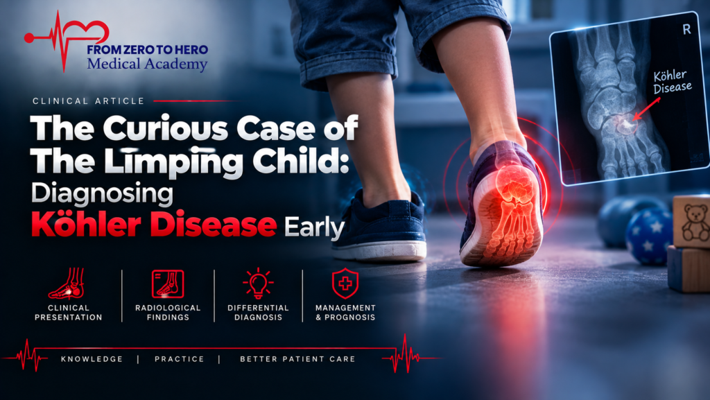
Köhler Disease: A Limping 4-Year-Old Boy with Foot Pain
Case Presentation
A 4-year-old boy presented to the Emergency Department with a limp and pain in the left foot lasting for two days.
History
- Mother denied any history of trauma or falls.
Examination
- Mild swelling and tenderness were noted over the dorsum of the left foot.
- Vital signs were all within normal limits.
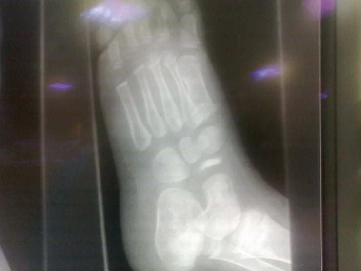
An X-ray of the affected foot was performed.
What Is the Diagnosis?
Upon careful review of the X-ray:
- The navicular bone appeared sclerotic, fragmented, hyperdense, small, and irregular.
This classic appearance leads to the diagnosis: Köhler Disease
Understanding Köhler Disease
What is Köhler Disease?
- A rare idiopathic condition caused by avascular necrosis of the navicular bone.
- Results in painful limp and localized tenderness over the navicular area.
Historical Note:
- First described by Dr. Alban Köhler, a German radiologist, in 1908.
Etiology:
- May follow minor trauma or occur without any prior injury.
Classification:
- Belongs to the group of crushing osteochondritis.
Epidemiology
- Prevalence: 2% of children.
- More common in boys (male to female ratio 5:1).
- Affects children aged 3–7 years.
- Usually unilateral, but bilateral involvement occurs in 25% of cases.
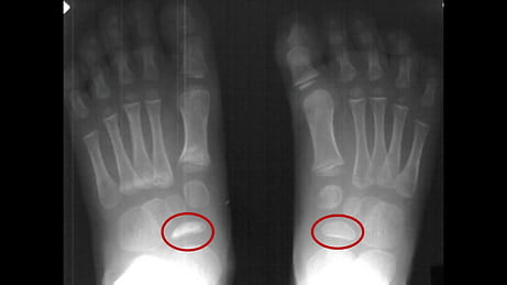
Radiographic Features
In Köhler Disease, the navicular bone typically appears:
- Sclerotic and hyperdense
- Fragmented and small
- Irregular compared to the normal navicular bone
Treatment
Conservative Management
- Pain control with rest and NSAIDs.
- Immobilization using a plaster cast (POP) in cases with severe symptoms.
- Orthopaedic follow-up is recommended.
Prognosis
- Excellent prognosis.
- Most cases resolve spontaneously within 6 to 48 months.
- No long-term disability reported.
Further Learning
Watch the full case discussion on YouTube
References
Key Takeaways
- Always consider Köhler Disease in young children with an unexplained limp and localized foot pain.
- Early imaging is crucial to avoid misdiagnosis.
- Conservative management leads to full recovery without long-term complications.
Stay tuned for more intriguing pediatric radiology cases!
FAQ
Q1: What is Köhler Disease?
Köhler Disease is avascular necrosis of the navicular bone in children, leading to pain and limping, often resolving spontaneously with conservative treatment.
Q2: What are the common radiographic features of Köhler Disease?
The navicular bone appears sclerotic, hyperdense, fragmented, small, and irregular on X-rays.
Q3: How is Köhler Disease treated?
Treatment is conservative, involving pain management, rest, possible immobilization in a cast, and follow-up with orthopaedics.

Ready To Take Your Clinical Learning Further?
Explore structured ECG and emergency medicine courses built around practical clinical knowledge, real cases, and confident decision-making.